 ultimate-guide
Receptor Binding & PK Profiling Support for Academic Labs
Table of Contents
- Why Academic Labs Need Specialized Support for Receptor Binding and PK Profiling
- Receptor-Ligand Binding Kinetics: What the Numbers Tell You
- Selecting a Receptor Binding Assay: A Practical Decision Guide
- Receptor Binding Assay Controls That Protect Your Data
- In Vitro ADMET Assays and the Plasma Protein Binding Assay in PK Profiling
- Interpreting Binding and Pharmacokinetic Metrics Together
- Study Design, Sample-Size Planning, and External Assay Support
- Step 1: Define the Estimand and the Decision
- Step 2: Set Concentration Ranges From Pilot Data
- Step 3: Choose Controls Before Replicates
- Step 4: Plan Replicates and Independent Runs
- Step 5: Size the Study With a Power Calculation
- Step 6: Decide What to Run In-House and What to Outsource
- Step 7: Pre-Register the Analysis Plan
- Budgeting, Timelines, and Documentation Standards for Academic Laboratories
- Frequently Asked Questions
Last Updated: October 18, 2026
Why Academic Labs Need Specialized Support for Receptor Binding and PK Profiling
A receptor binding study lives or dies on compound quality. At Globo Research Peptides, we supply research-grade peptides with verified ≥99% HPLC purity and a full Certificate of Analysis for every batch, because support for in vitro receptor binding and pharmacokinetic profiling studies for academic laboratories starts with material you can trust before the first assay runs.
Academic labs face a specific squeeze: short grant cycles, core facilities booked weeks out, and a single failed batch that can push a publication back a full semester. Outsourcing selected assays or sourcing better-characterized compounds often decides whether a study finishes on time.
Receptor-Ligand Binding Kinetics: What the Numbers Tell You
Receptor-ligand binding kinetics describe how fast a ligand associates with and dissociates from its receptor, and how tightly it binds at equilibrium. Three constants carry that story: kon, koff, and Kd.
- kon (association rate) reflects how quickly the complex forms, usually reported in M⁻¹s⁻¹.
- koff (dissociation rate) reflects how quickly it falls apart, in s⁻¹.
- Kd (equilibrium dissociation constant) is the ratio koff/kon and defines binding affinity.
Reading kon, koff, and Kd Together
Two ligands can share an identical Kd and behave nothing alike. A fast-on, fast-off compound reaches equilibrium quickly and washes out readily; a slow-on, slow-off compound may take hours to plateau but stays bound long after free ligand is gone.
That distinction matters when you plan wash steps, incubation times, and sampling windows. Design a wash protocol around Kd alone and a slow-dissociating ligand will look artificially potent.
Selecting a Receptor Binding Assay: A Practical Decision Guide
The right receptor binding assay depends on whether you need equilibrium affinity, kinetic rates, or target engagement in a complex matrix. Most academic labs lack every platform, so start with the question and the equipment you can actually access, then narrow to a method.
| Assay Type | Measures | Best For | Main Limitation |
|---|---|---|---|
| Radioligand filtration | Kd, Bmax | Membrane preparations, high-throughput screens | Radioactive waste handling and licensing |
| Surface plasmon resonance | kon, koff, Kd | Kinetic profiling, label-free work | Immobilization can alter receptor conformation |
| Fluorescence polarization | Kd, competition | Small-molecule screening | Interference from fluorescent compounds |
| Cell-based binding | Target engagement | Native receptor context | Lower precision, higher variability |
| Equilibrium dialysis (for free fraction) | Fraction unbound | Linking binding to exposure | Slow equilibration, compound adsorption |
A Four-Question Decision Framework
Work through these in order. The first question that produces a hard constraint usually settles the method.
- What output does the paper or grant actually require? If reviewers need kon and koff, only a kinetic method (SPR, or radioligand association/dissociation curves at multiple time points) will do. For Kd and Bmax on a membrane preparation, filtration is the direct route; for target engagement in cells, a cell-based format is the honest one.
- What equipment can you reach this quarter? An SPR instrument booked through a core facility may have a two- to six-week wait; a filtration manifold and plate reader are usually available same-week. Match the method to the access window, not the ideal experiment.
- What is the compound's detection profile? Fluorescent compounds interfere with fluorescence polarization; lipophilic compounds adsorb to filtration and dialysis membranes alike. If the compound is problematic on one platform, that platform is disqualified regardless of its other merits.
- What throughput does the study need? A single Kd determination tolerates a slow, precise method; a 200-compound series does not. Filtration and fluorescence polarization scale to 96- and 384-well plates; SPR is low-throughput, high-information.
Matching Method to Target Class
- G-protein-coupled receptors are commonly run as radioligand filtration assays on membrane preparations, because the receptor is abundant and the radioligand toolbox is mature. SPR is harder here because solubilized or immobilized GPCRs often lose native conformation.
- Soluble receptors and enzyme targets are good candidates for SPR, where immobilization is more forgiving and kinetic constants are the point.
- Nuclear receptors and other intracellular targets often call for cell-based or fluorescence-based formats, because the native context matters and membrane preparations do not capture it.
When to Combine Methods
The strongest manuscripts rarely rely on one platform. A common pattern is a filtration saturation curve to establish Kd and Bmax, followed by SPR on a subset of compounds to extract kon and koff, with a cell-based assay to confirm engagement in a native context. The combination costs more instrument time but answers the reviewer question a single method cannot: does the binding number mean what we think it means?
A Note on Outsourcing the Method You Cannot Run
If the decision framework lands on a method your lab cannot access in time, external assay support is the practical answer. Ask a provider which platform they will use, what controls they run, and whether they will return raw curves rather than only fitted constants.
Receptor Binding Assay Controls That Protect Your Data
Receptor binding assay controls separate real signal from artifacts, and reviewers increasingly ask for them by name. Build them into the protocol before the first plate runs, not after a revision request.
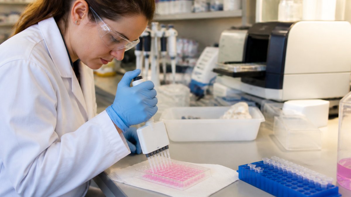
The non-negotiable set:
- Total binding wells with ligand and receptor only
- Non-specific binding wells with excess cold competitor
- Nonspecific filter binding wells without receptor, to catch ligand sticking to the matrix
A common mistake is treating non-specific binding as a formality. High filter adsorption inflates apparent affinity and produces flat, unusable curves.
In Vitro ADMET Assays and the Plasma Protein Binding Assay in PK Profiling
In vitro ADMET assays predict absorption, distribution, metabolism, excretion, and toxicity before an animal study is funded. They include permeability screens, metabolic stability in liver microsomes, CYP inhibition panels, and plasma protein binding.
Where the Plasma Protein Binding Assay Fits
The plasma protein binding assay measures what fraction of a compound is free in plasma. Only unbound drug reaches the receptor, so a strongly bound compound can look potent in buffer and inert in circulation.
Run this assay early. It reframes every downstream number: a modest shift in free fraction changes the exposure you need to reach target engagement.
Interpreting Binding and Pharmacokinetic Metrics Together
Binding affinity and pharmacokinetics answer different halves of the same question, and reading them separately produces confident, wrong conclusions.
Connect the two through free drug exposure. Take total plasma concentration, correct it by free fraction from the plasma protein binding assay, and compare that free concentration to your Kd.
Key PK parameters to place alongside binding data:
- Cmax and AUC describe exposure magnitude and duration
- Clearance and half-life describe how long exposure persists
- Bioavailability describes how much reaches circulation
A compound with sub-nanomolar Kd and rapid clearance can fail where a weaker binder with sustained exposure succeeds.
Study Design, Sample-Size Planning, and External Assay Support
Sound study design sets sample size before data collection, not after. For binding experiments, replicate wells across at least three independent runs and use a power calculation based on pilot variability rather than habit.
Step 1: Define the Estimand and the Decision
Write down the number the study must produce and the decision it feeds. "A Kd with a confidence interval tight enough to rank three compounds" is a design specification. "Characterize binding" is not. The estimand determines whether you need a full saturation curve, a single-point screen, or a kinetic run.
Step 2: Set Concentration Ranges From Pilot Data
A saturation curve should span roughly 0.1× to 10× the expected Kd, with enough points around the Kd to define the inflection. If the Kd is unknown, run a broad pilot first, then narrow. For competition experiments, center the series on the expected IC50 with at least two points on each side of the inflection; a curve with no points near the inflection yields a wide confidence interval no matter how many replicates you run.
Step 3: Choose Controls Before Replicates
Controls are cheaper than replicates and catch more errors. The non-negotiable set:
- Total binding wells with ligand and receptor only
- Non-specific binding wells with excess cold competitor
- Nonspecific filter or plate binding wells without receptor
If the positive control does not reproduce its published Kd within your assay's variability, stop and fix the assay before running the study compounds.
Step 4: Plan Replicates and Independent Runs
Technical replicates (multiple wells of the same sample on one plate) estimate within-plate variability. Independent runs (the same experiment repeated on different days, with fresh dilutions) estimate the variability that actually matters for a manuscript. A common pattern is triplicate wells across at least three independent runs, with the reported Kd being the mean of the run-level fits rather than a single pooled fit.
Step 5: Size the Study With a Power Calculation
Use pilot variability, not textbook defaults. If the pilot standard deviation is large relative to the effect you need to detect, more replicates will not rescue a poorly designed curve, fix the curve first. For the PK arm, base sample numbers on pilot-cohort variability and state the assumed effect size in the methods.
Step 6: Decide What to Run In-House and What to Outsource
External assay support fills gaps academic labs cannot always close in-house: kinetic runs on an SPR instrument, ADME panels, or validated binding assays on short notice. The NIH Guidelines for Research Involving Recombinant or Synthetic Nucleic Acid Molecules and institutional IACUC and IRB review processes set the compliance frame for the work these assays support, so confirm documentation requirements before shipping samples.
When evaluating a provider, ask for raw data, method descriptions, and instrument calibration records. A report without those is hard to defend in supplementary methods. A short scoping checklist helps:
- Which platform will be used, and why that platform for this target?
- What controls are run, and are they reported alongside the study data?
- Will raw curves and fit parameters both be returned?
Step 7: Pre-Register the Analysis Plan
Write the analysis plan before unblinding: which model fits the curve, how outliers are handled, and which run-level statistic is reported. A pre-registered plan is the cheapest reproducibility safeguard available and strengthens a grant renewal as much as a manuscript.
Budgeting, Timelines, and Documentation Standards for Academic Laboratories
Budget and timeline planning for binding and PK work follows a predictable shape, and most overruns trace to two causes: assay rework and compound resupply.
| Phase | Typical Duration | Main Cost Driver |
|---|---|---|
| Compound sourcing and QC | 1-3 weeks | Purity verification, COA review |
| Assay development and validation | 2-6 weeks | Instrument time, pilot runs |
| Sample analysis | 2-4 weeks | Replicates, plate count |
| Data analysis and reporting | 1-2 weeks | Statistical support |
Documentation standards deserve the same attention. The FDA guidance on bioanalytical method validation sets expectations for the assay documentation that supports regulatory-facing work, and the same discipline strengthens a grant renewal.
Funding routes vary: internal pilot funds, core facility subsidies, and shared instrumentation grants all cover pieces of this work.
For peptide work specifically, PubChem's compound database is a useful cross-check for identity and published reference data before you commit a batch to a study.
Frequently Asked Questions
What is an in vitro receptor-binding assay?
An in vitro receptor-binding assay measures how a ligand interacts with a receptor outside a living organism, typically using membrane preparations or purified receptor protein. Radiolabeled or fluorescent ligands compete with test compounds for binding sites, and the resulting data yield binding affinity (Kd) and kinetics (kon, koff). These assays are a standard first step in preclinical research because they establish target engagement before you commit to cell-based or in vivo work. The quality of your receptor binding data depends heavily on assay controls and compound purity.
How do researchers measure receptor-binding affinity and kinetics?
Affinity is usually derived from saturation or competition binding experiments, where you fit the data to a binding model to obtain Kd. Kinetics require time-resolved measurements: surface plasmon resonance or stopped-flow fluorescence can capture association (kon) and dissociation (koff) rates directly. For academic laboratories, radioligand filtration assays remain common because they are cost-effective and well documented. Whichever method you choose, run replicates and include a known reference ligand so you can benchmark your receptor-ligand binding kinetics against published values.
Which in vitro assays are used to profile pharmacokinetic properties?
Early PK profiling typically combines metabolic stability assays (microsomes or hepatocytes), permeability assays such as Caco-2, plasma protein binding assays, and solubility screens. Together these form the core of in vitro ADMET assays that predict absorption, distribution, metabolism, and excretion before dosing. A plasma protein binding assay tells you the free fraction available to engage your receptor, which directly affects how you interpret exposure metrics like Cmax and AUC. Running these assays early helps academic groups prioritize compounds and design better in vivo studies.
What controls should be included in receptor-binding studies?
At minimum, include a total binding well, a nonspecific binding well with excess unlabeled ligand, and a filter or membrane background control. A reference competitor with known affinity validates that your receptor preparation behaves as expected. Negative controls, such as wells without receptor, catch plate-binding artifacts. Receptor binding assay controls also should cover vehicle effects and temperature equilibration. Skipping these controls is one of the most common reasons binding data fail to reproduce across labs, so build them into your protocol from the start.
How can academic laboratories interpret receptor-binding and pharmacokinetic data together?
Binding data tell you how tightly a compound engages its target; PK data tell you whether free compound reaches that target at a useful concentration and for how long. Compare your Kd with free-drug exposure (Cmax and AUC adjusted for plasma protein binding) to judge whether target engagement is plausible in vivo. If free concentrations sit well below Kd, the compound is unlikely to occupy enough receptors regardless of how good the binding affinity looks. Interpreting both datasets together prevents over-optimistic conclusions from binding assays alone.
What information should researchers provide when requesting external assay support?
Share your receptor source and preparation method, the ligand and radioligand you plan to use, expected affinity ranges, buffer conditions, and any published protocols you are following. Specify your study design, including replicate numbers and controls, and state the data format you need for analysis. If you are sourcing peptides, request a Certificate of Analysis with purity and identity data so the assay provider can confirm compound quality. Clear inputs reduce turnaround time and prevent costly repeats in receptor binding and PK profiling projects.
Academic binding and PK studies fail quietly when compound quality and documentation are treated as an afterthought. Globo Research Peptides supplies research-grade peptides with verified ≥99% HPLC purity, full Certificate of Analysis documentation for every batch, and rigorous third-party testing, plus a special-order program for compounds outside standard inventory. Get started with Globo Research Peptides and build your next study on material you can document.