 listicle
5 Best Practices for Peptide Mapping Protocols
Table of Contents
- Why Peptide Mapping Protocols Succeed or Fail
- 1. Lock Down Peptide Mapping Sample Preparation Before Anything Else
- 2. Build a Peptide Mapping Workflow Around Selective Cleavage
- 3. Separate Peptides by Liquid Chromatography Before Detection
- 4. Push Peptide Mapping Sequence Coverage Toward Completeness
- 5. Treat Peptide Mapping Data Analysis as a Controlled Process
- Troubleshooting: Symptoms, Causes, and Fixes
- Conclusion: Turning Five Practices Into a Repeatable Protocol
- Frequently Asked Questions
Last Updated: October 12, 2026
Why Peptide Mapping Protocols Succeed or Fail
Peptide mapping is the analytical process of cleaving a protein into peptides and separating them by liquid chromatography so each fragment can be identified by mass spectrometry. It confirms protein identity, sequence, and post-translational modifications.
The stakes are concrete. A 2025 review in PubMed Central on peptide mapping for sequence confirmation describes peptide mapping as a core method for confirming therapeutic protein identity in batch-release testing, which means a sloppy protocol has regulatory consequences, not just academic ones.
1. Lock Down Peptide Mapping Sample Preparation Before Anything Else
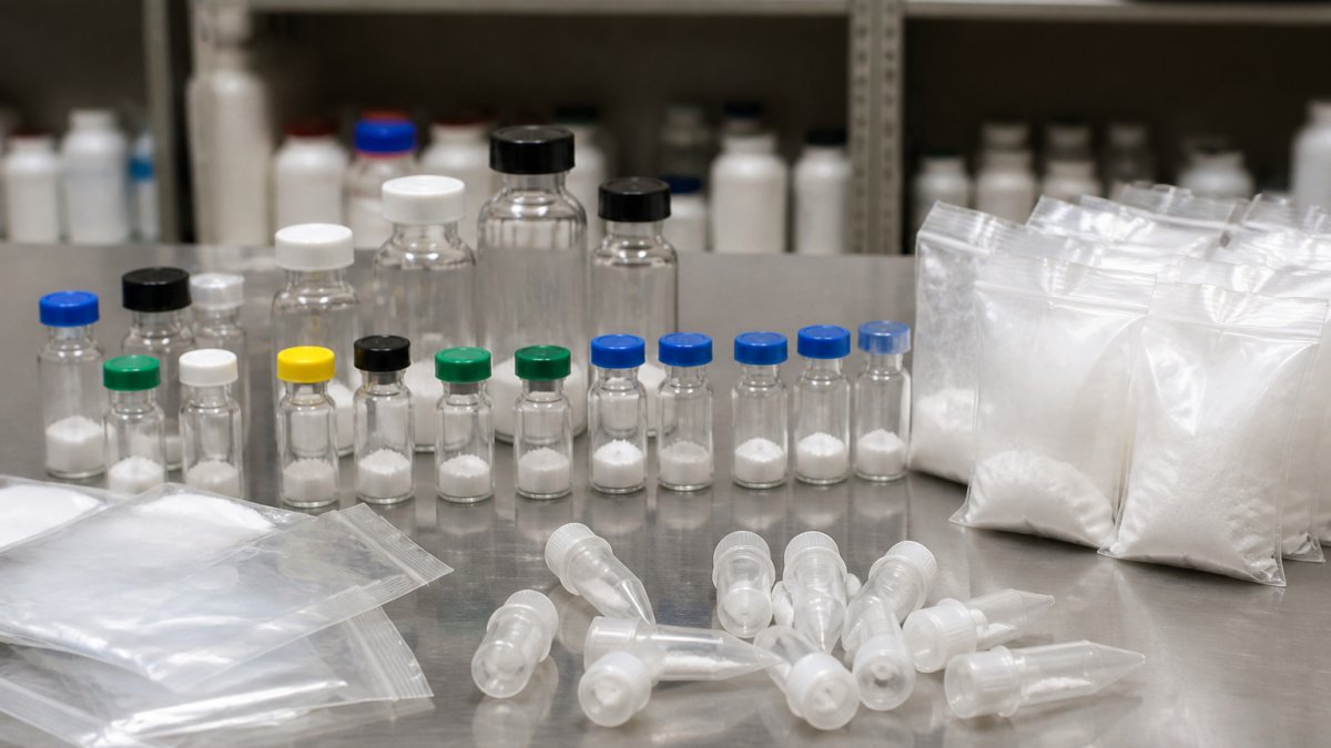
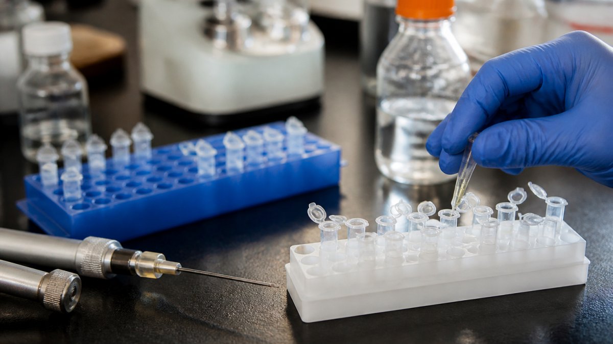
Peptide mapping sample preparation is where reproducibility is won or lost. As Chromatography Online's overview of validation and peptide mapping notes, peptide-map generation is a multistep process beginning with protein isolation and purification.
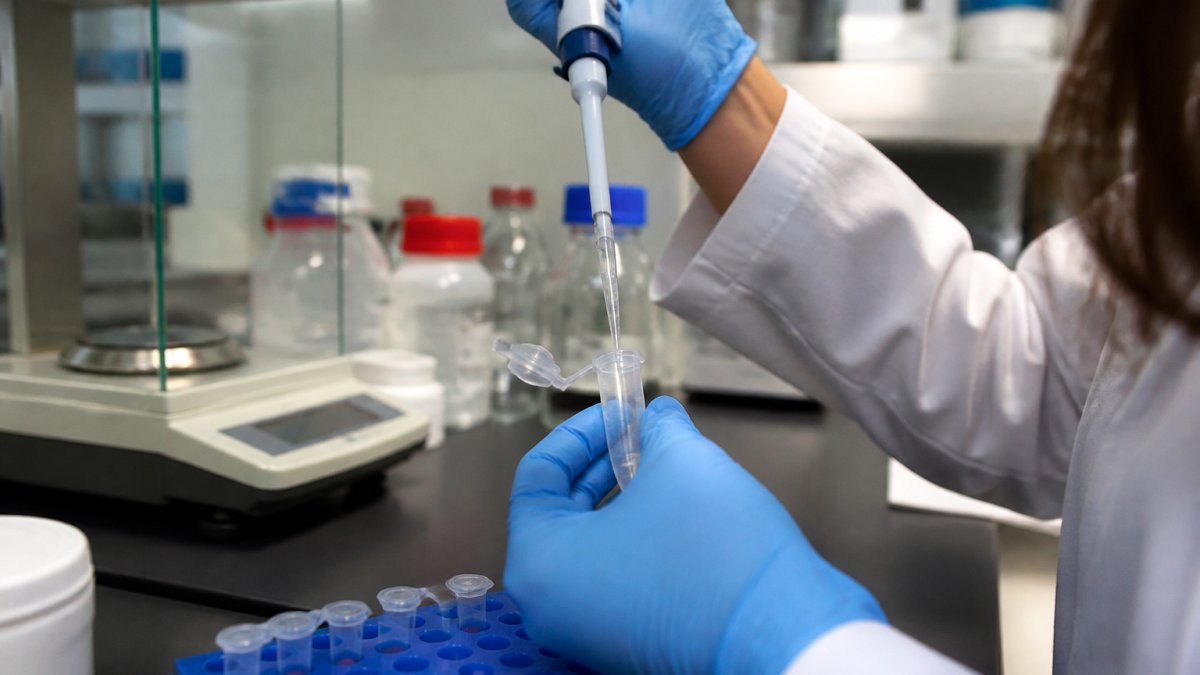
[IMAGE: A scientist in gloves and lab coat pipetting a protein sample into a small microcentrifuge tube on a clean benchtop, with an analytical balance and reagent bottles softly blurred in the background | section:1.
Denaturation, Reduction, and Alkylation: The Steps That Decide Your Map
Denaturation unfolds the protein so the protease can reach cleavage sites. Reduction breaks disulfide bonds. Alkylation caps the freed cysteines so they cannot re-form bonds and scramble your map.
A common mistake is rushing the alkylation quench. Residual alkylating agent modifies unintended residues and creates artifact peaks that look like real post-translational modifications, so your analysis flags a modification that does not exist.
Sample Matrix and Artifact Risks That Quietly Corrupt Results
Sample matrix components, salts, detergents, and buffer additives, interfere with both digestion and LC-MS detection. A protocol that works on a clean reference standard can fail on a formulated sample because the matrix changes enzyme activity and ionization efficiency. Desalting or buffer exchange before digestion is not optional for complex matrices.
2. Build a Peptide Mapping Workflow Around Selective Cleavage
A peptide mapping workflow is only as good as its cleavage step. Selective cleavage determines peptide size, ionization behavior, and which regions of the sequence you can observe. Choose the protease and digestion conditions deliberately, then lock them into your protocol.
Protease Selection: Trypsin and Alternatives
Trypsin remains the default because it cleaves at the C-terminal side of lysine and arginine, producing peptides in a mass range that suits LC-MS/MS. Alternatives exist for specific problems: other proteases handle missed cleavage hotspots, hydrophobic regions, or proteins with unusual residue distributions. Trypsin quality matters as much as protease choice. Reagents such as RapiZyme Trypsin have been featured in Waters' LC-UV-MS peptide mapping application note for biotherapeutic workflows, where reagent consistency directly affects map reproducibility.
Digestion Conditions: Time, Temperature, and Enzyme-to-Substrate Ratio
Three variables decide digestion completeness: time, temperature, and enzyme-to-substrate ratio. Under-digest and you get missed cleavages that inflate peptide size and hurt sequence coverage; over-digest and you get nonspecific cleavage and artifact peptides. Determine the right values experimentally with a small matrix study rather than copying a published protocol.
| Variable | Symptom if Too Low | Symptom if Too High |
|---|---|---|
| Digestion time | Missed cleavages, large peptides | Nonspecific cleavage, artifacts |
| Temperature | Incomplete digestion | Enzyme autolysis, artifacts |
| Enzyme-to-substrate ratio | Incomplete digestion | Over-digestion, cost waste |
3. Separate Peptides by Liquid Chromatography Before Detection
Liquid chromatography separates; mass spectrometry identifies. Reversed-phase LC with a shallow gradient resolves peptides by hydrophobicity, and gradient slope decides how many peptides you can distinguish. Too steep co-elutes peptides and buries low-abundance fragments; too shallow stretches run time without adding resolution.
Gradient Slope and Peak Capacity: The Math That Decides Resolution
Peak capacity is the practical ceiling on how many peptides you can resolve in a single run, and it scales with gradient time and inversely with peak width.
Gradient slope is expressed as percent organic per column volume, not percent per minute, because the same slope on a longer column produces a different separation. When you change column length or particle size, recalculate slope in column volumes first, the most common reason a method that transfers cleanly between instruments still produces a different map.
Column Selection Trade-Offs You Should Document
Smaller particles and longer columns improve peak capacity but raise backpressure and shorten column life. Match the column to your instrument's pressure limit and throughput needs. Decision criteria worth writing into your protocol:
| Objective | Column Direction | Trade-Off to Accept |
|---|---|---|
| Maximum resolution for a complex digest | Longer column, smaller particles | Higher backpressure, shorter column life, longer runs |
| High-throughput batch release | Shorter column, larger particles | Lower peak capacity, more co-elution risk |
| Hydrophilic peptide retention | Lower organic starting condition, ion-pairing agent | Longer equilibration, possible ion suppression |
| Hydrophobic peptide recovery | Higher organic wash, steeper late gradient | Possible carryover into next injection |
Method development here is incremental: change one variable, measure resolution and peak shape, keep what improves reproducibility. Record the column lot, gradient in column volumes, flow rate, and column temperature for every method version, those four values make a method transferable.
Carryover, Equilibration, and the Injection-to-Injection Drift Nobody Logs
Carryover from hydrophobic peptides shows up as a ghost peak in the blank and a falsely elevated modification call in the next sample. Run a blank after every high-abundance sample during method development; if carryover appears, add a wash step with a higher organic percentage than the final gradient condition. Equilibration volume matters too: under-equilibrating shifts retention times run to run and makes acceptance criteria meaningless.
System Suitability Before You Trust a Batch
Before running study samples, inject a system suitability standard and confirm resolution between a critical pair, peak asymmetry, and retention time reproducibility all meet pre-set limits. If the standard fails, the batch fails, do not proceed and hope. This separates a method that survives audit from one that produced a good-looking map once.
4. Push Peptide Mapping Sequence Coverage Toward Completeness
Peptide mapping sequence coverage is the percentage of the protein sequence you can observe. High coverage is the difference between confirming identity and guessing at it. Low coverage usually traces to three causes: missed cleavages that leave a region in an unmeasurable peptide, poor ionization of specific peptides, and peptides lost during sample preparation. Diagnose which one is operating before you change a variable.
Set a Coverage Target Before You Run
Coverage expectations should be defined by the analytical objective, not by what the last run produced. Identity confirmation typically targets near-complete coverage, accepting that a few short or highly hydrophilic peptides may be hard to retain. For monitoring a specific modification, the target is complete coverage of the region around that site, even if overall coverage is lower.
Diagnose the Cause Before You Change the Method
Use the pattern of missing peptides to narrow the cause:
| Coverage Gap Pattern | Likely Cause | Diagnostic Step |
|---|---|---|
| Missing peptides cluster around Lys/Arg sites | Missed cleavage | Check enzyme-to-substrate ratio and digestion time |
| Missing peptides are short and hydrophilic | Poor retention on reversed-phase | Inject a hydrophilic standard; adjust gradient start |
| Missing peptides are large and hydrophobic | Poor ionization or solubility | Check for precipitation; try a different protease |
| Coverage drops only in formulated samples | Matrix interference | Desalt or buffer-exchange before digestion |
| Coverage varies run to run | Sample prep or LC variability | Run a reference material alongside study samples |
Change one variable at a time, re-run, and compare the coverage map against the previous run.
Close Gaps Systematically, Not by Trial and Error
If a region is consistently missing, try a second protease with different cleavage specificity to generate an overlapping peptide covering the gap. Adjust the LC gradient to retain hydrophilic peptides, or use a different ionization mode for poorly ionizing peptides. For regions that resist every approach, consider whether the gap is inherent, a very short peptide between two cleavage sites may simply be below the detection limit, a coverage limit worth documenting rather than chasing.
Controls and Acceptance Criteria That Make Coverage Defensible
A coverage number without controls is not evidence. Build these into every batch:
- A blank injection to confirm no carryover is inflating or masking peaks
- A reference material with a known map to confirm the system is performing as expected
- Replicate injections to establish run-to-run coverage variability
Document which regions are hard to cover and why, that record becomes your method's robustness story. A protocol that acknowledges its coverage limits is stronger in review than one claiming completeness it cannot demonstrate.
5. Treat Peptide Mapping Data Analysis as a Controlled Process
Peptide mapping data analysis is a controlled step with defined acceptance criteria. Set these before you run samples, not after you see the data:
- Minimum sequence coverage threshold for your protein class
- Mass tolerance for peptide identification
- Criteria for accepting or rejecting a post-translational modification call
Apply the same search parameters, reference sequence, and acceptance criteria to every batch. Reproducibility across runs matters, not the quality of a single map. When you change software versions or search parameters, re-analyze a reference material and confirm the results still match.
Troubleshooting: Symptoms, Causes, and Fixes
Match your symptom to the likely cause before changing anything else, changing multiple variables at once destroys your ability to learn what fixed the problem.
| Symptom | Likely Cause | Fix |
|---|---|---|
| Low sequence coverage | Missed cleavages, lost hydrophilic peptides | Optimize digestion; adjust gradient |
| Extra peaks not in reference | Alkylation artifacts, nonspecific cleavage | Quench alkylation fully; reduce enzyme ratio |
| Poor run-to-run reproducibility | Sample prep variability, column degradation | Standardize prep; replace column |
| Weak signal early in run | Salt or detergent interference | Desalt or buffer exchange before digestion |
| Missing modification calls | Low peptide abundance, tight mass tolerance | Enrich modified peptides; widen tolerance |
Conclusion: Turning Five Practices Into a Repeatable Protocol
The hard part of peptide mapping is not any single step; it is holding five steps consistent across every batch and every analyst. Most labs get the science right and the process wrong. Globo Research Peptides provides verified ≥99% HPLC purity and a full Certificate of Analysis for every batch, so your reference material starts from a documented baseline.
Frequently Asked Questions
What is peptide mapping used for?
Peptide mapping is used to confirm protein identity, verify amino acid sequence, and characterize therapeutic proteins and biotherapeutics. It supports sequence confirmation, detection of post-translational modifications, and batch-release testing. Because the method breaks a protein into peptides through enzymatic digestion and then separates and detects them by liquid chromatography and mass spectrometry, it provides a detailed fingerprint that can be compared against a reference material. This makes peptide mapping a core tool in product characterization and consistency testing across development and quality control.
How does peptide mapping confirm a protein sequence?
The workflow starts with protein isolation and purification, followed by denaturation, reduction, and alkylation to unfold the protein and protect cysteine residues. A protease such as trypsin then cleaves the chain into peptides at specific sites. Those peptides are separated by liquid chromatography and analyzed by mass spectrometry, often LC-MS/MS. Software matches the observed peptide masses and fragmentation patterns to the expected sequence, and the percentage of the sequence covered by detected peptides, known as sequence coverage, indicates how thoroughly the protein identity has been confirmed.
What causes missed cleavages in peptide mapping?
Missed cleavages usually trace back to digestion conditions or sample preparation. Common causes include insufficient denaturation, incomplete reduction and alkylation, too little protease relative to substrate, shortened digestion time, or a sample matrix that interferes with enzyme activity. Trypsin also will not cleave efficiently next to certain residues or when the target site is buried in folded structure. Increasing enzyme-to-substrate ratio, extending digestion time, or switching to a different protease can reduce missed cleavages, but each change should be evaluated against a reference material to confirm it improves rather than degrades the peptide map.
How can you improve sequence coverage in peptide mapping?
Sequence coverage improves when more of the protein is converted into detectable peptides. Practical steps include optimizing digestion conditions, using a protease with appropriate specificity, and, in some cases, combining results from more than one enzyme to cover regions that trypsin alone misses. Adjusting liquid chromatography gradients to resolve hydrophilic peptides that elute early can also help. The goal is to maximize the proportion of the sequence that is observed while keeping the method reproducible, so any change should be verified with a reference material and documented as part of method development.
How do you assess the reproducibility of a peptide mapping protocol?
Reproducibility is assessed by repeating the full workflow, from sample preparation through data analysis, and comparing the resulting peptide maps. Key metrics include retention time consistency, relative peak area variability, and the number of peptides identified across replicates. Running the protocol on a reference material alongside unknown samples provides a control that flags drift in digestion, chromatography, or detection. Acceptable ranges for these metrics should be defined in advance as part of method validation, and any deviation should trigger a review of the specific step, such as digestion time or gradient performance, that most likely caused it.